
Journal Article
Validation of Smartphone-Based Retinal Photography for Diabetic Retinopathy Screening
MII Ret Cam featured on Cover Page
Ophthalmic Surg Lasers Imaging Retina · 2019
View PDF / PubMedDiscover the published scientific evidence behind MII Ret Cam technologies. This collection features peer-reviewed journal articles authored by ophthalmologists from around the world. Access the full-text PDF, journal website, or PubMed listing with a single click.
Ophthalmic Surg Lasers Imaging Retina · 2019
View PDF / PubMed
J Glaucoma · 2019
View PDF / PubMed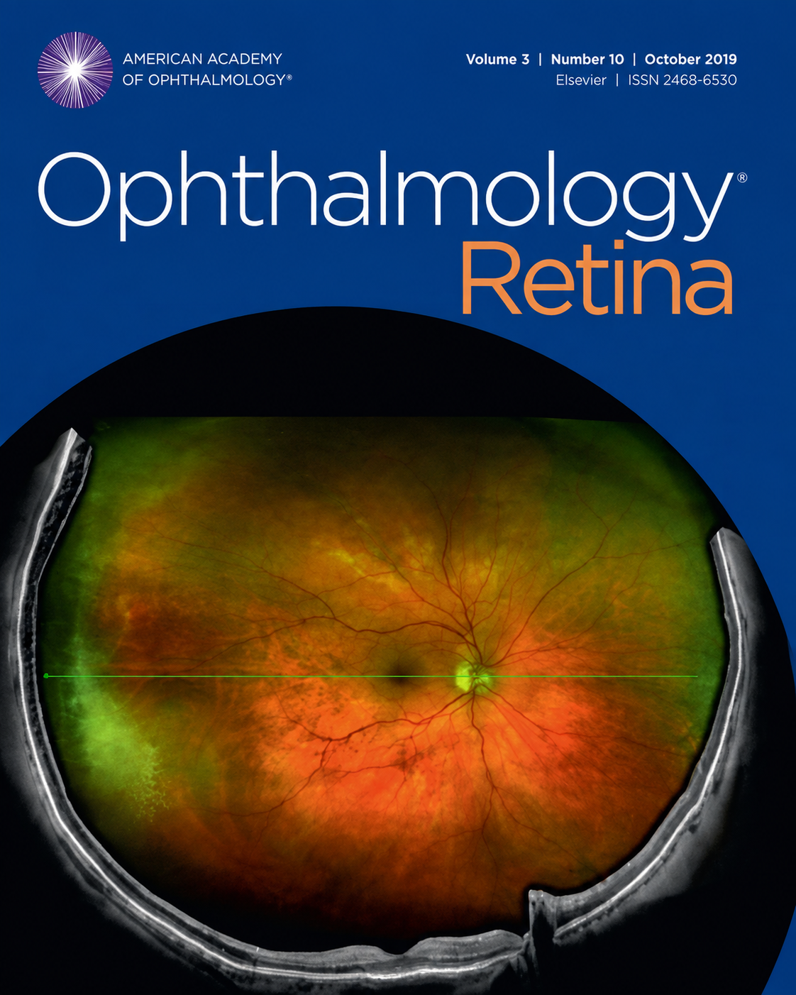
Ophthalmology · 2019
View PDF / PubMed
JAMA Ophthalmology · 2023
View PDF / PubMed
Eye · 2018
View PDF / PubMed
Eye · 2020
View PDF / PubMed

European Journal of Ophthalmology · 2016
View PDF / PubMed
Eye (Lond) · 2020
View PDF / PubMed
J Fr Ophtalmol · 2020
View PDF / PubMed
Journal of Medicine, Surgery, and Public Health · 2024
View PDF / PubMed
Indian J Ophthalmol · 2019
View PDF / PubMed
Indian J Ophthalmol · 2019
View PDF / PubMed
Indian J Ophthalmol · 2019
View PDF / PubMed
Indian J Ophthalmol · 2023
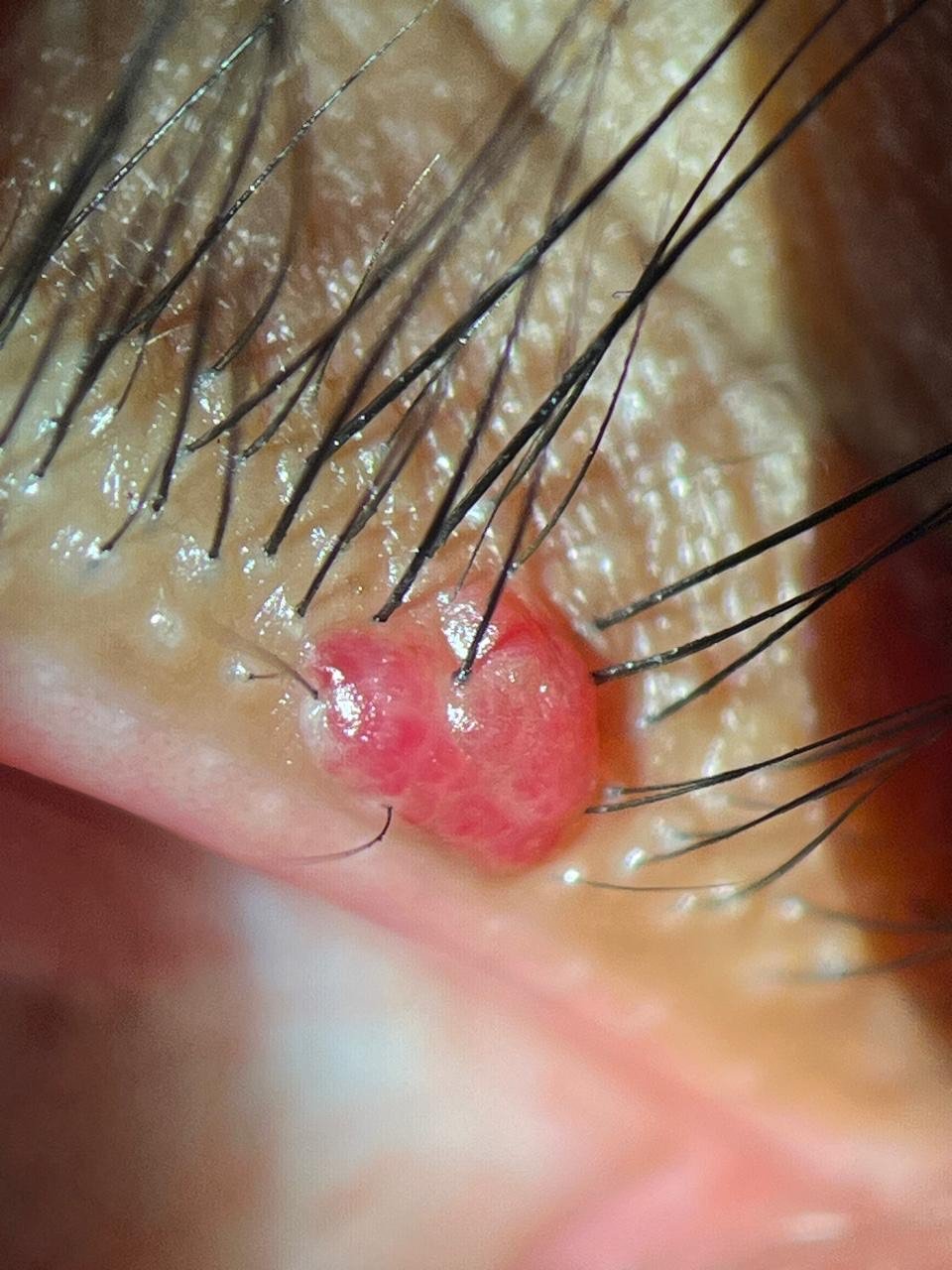
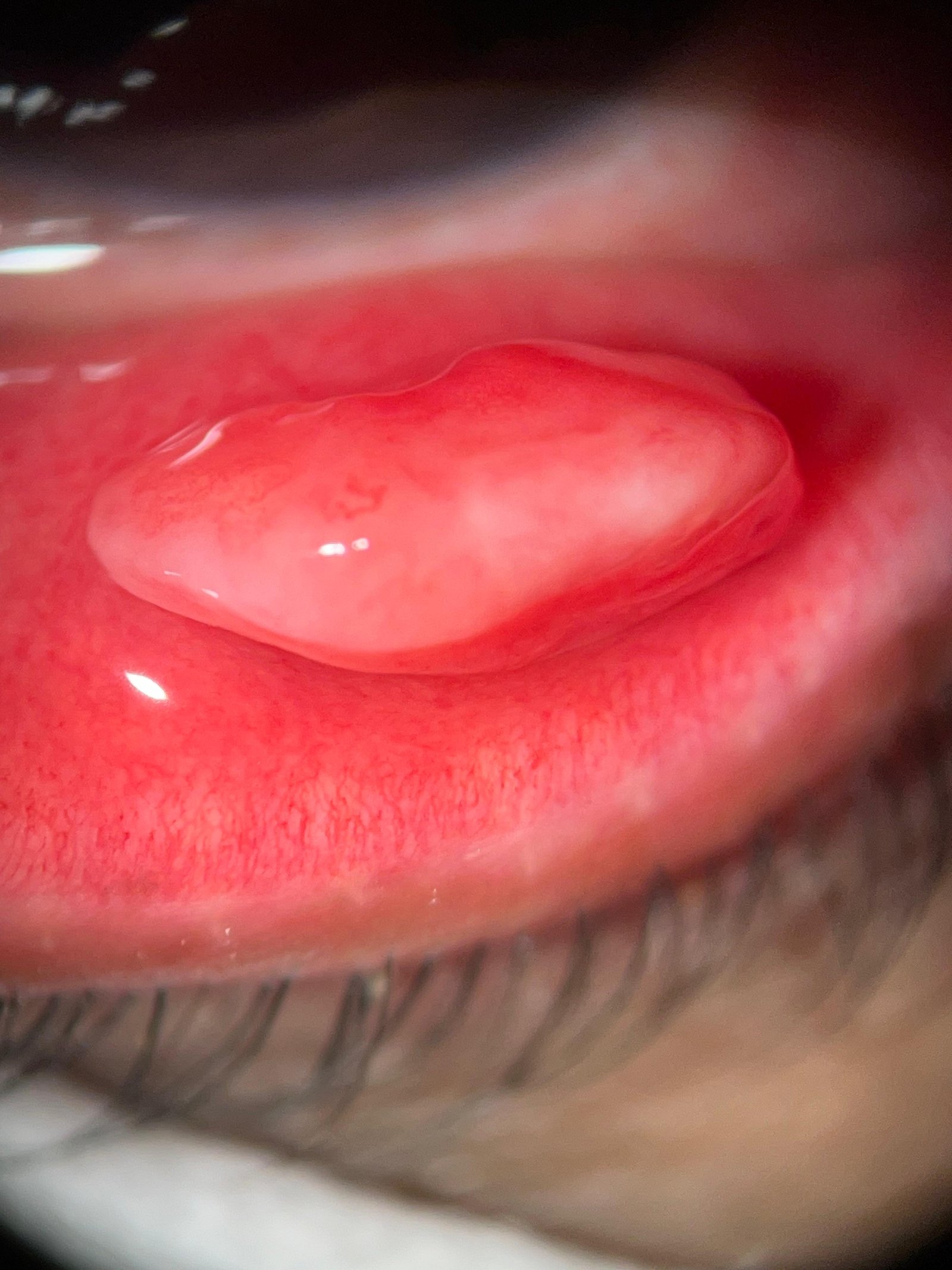
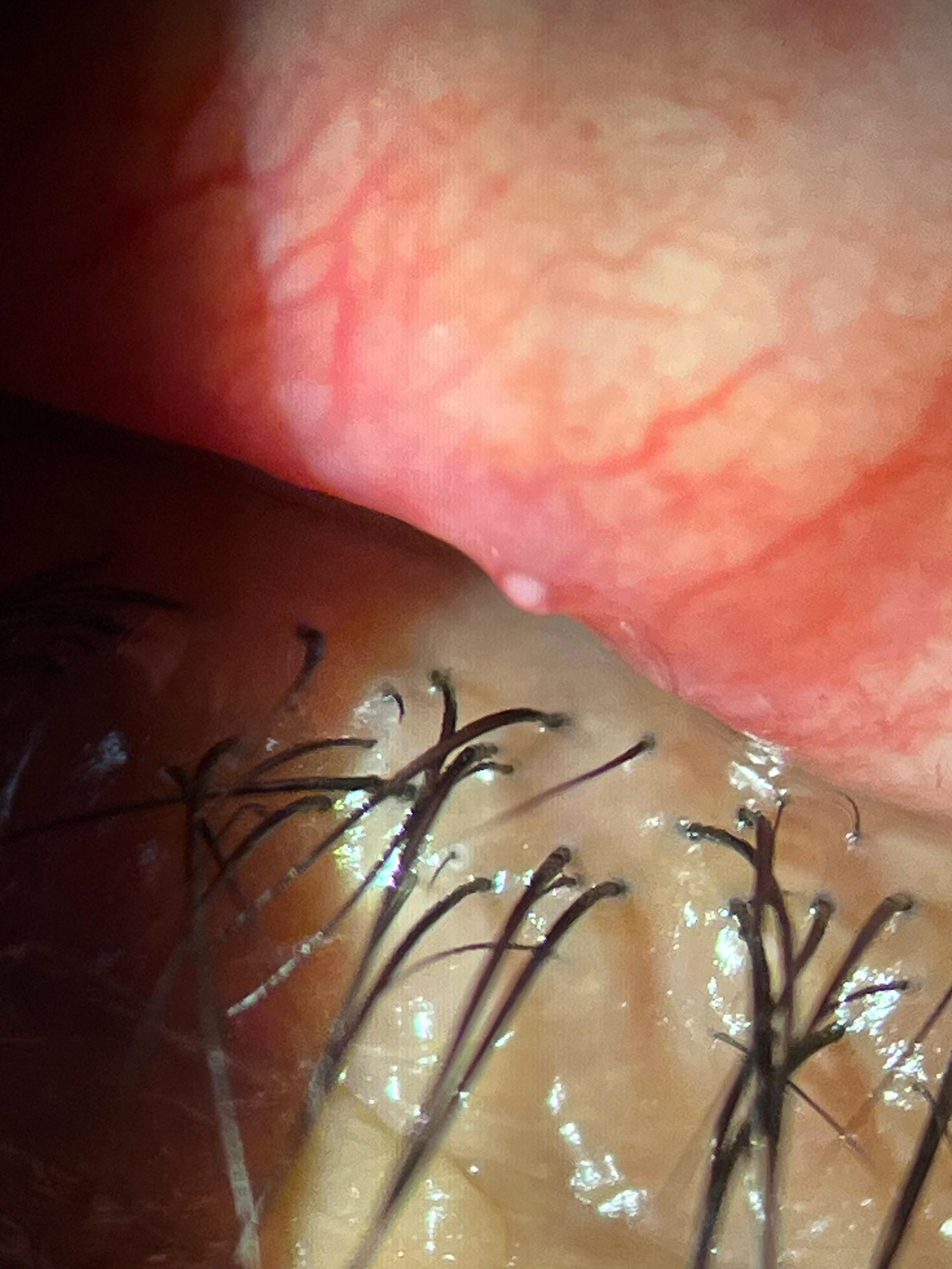
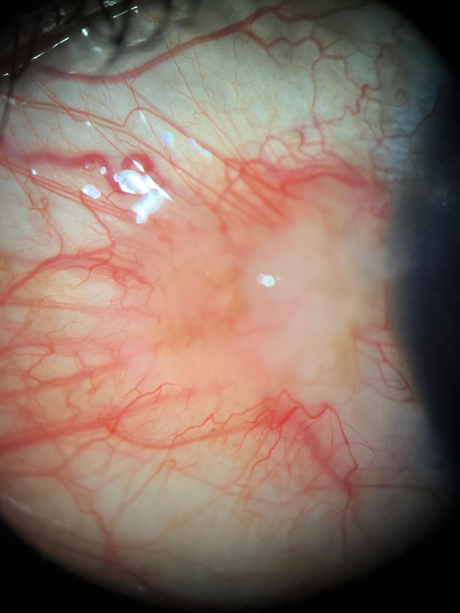
View PDF / PubMedA look at MII devices in everyday use at eye clinics and hospitals nationwide.
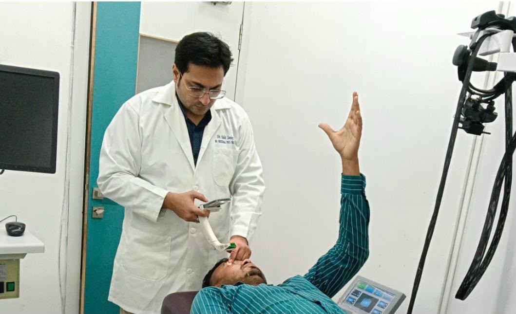
Dr. Rohit Laul
Nashik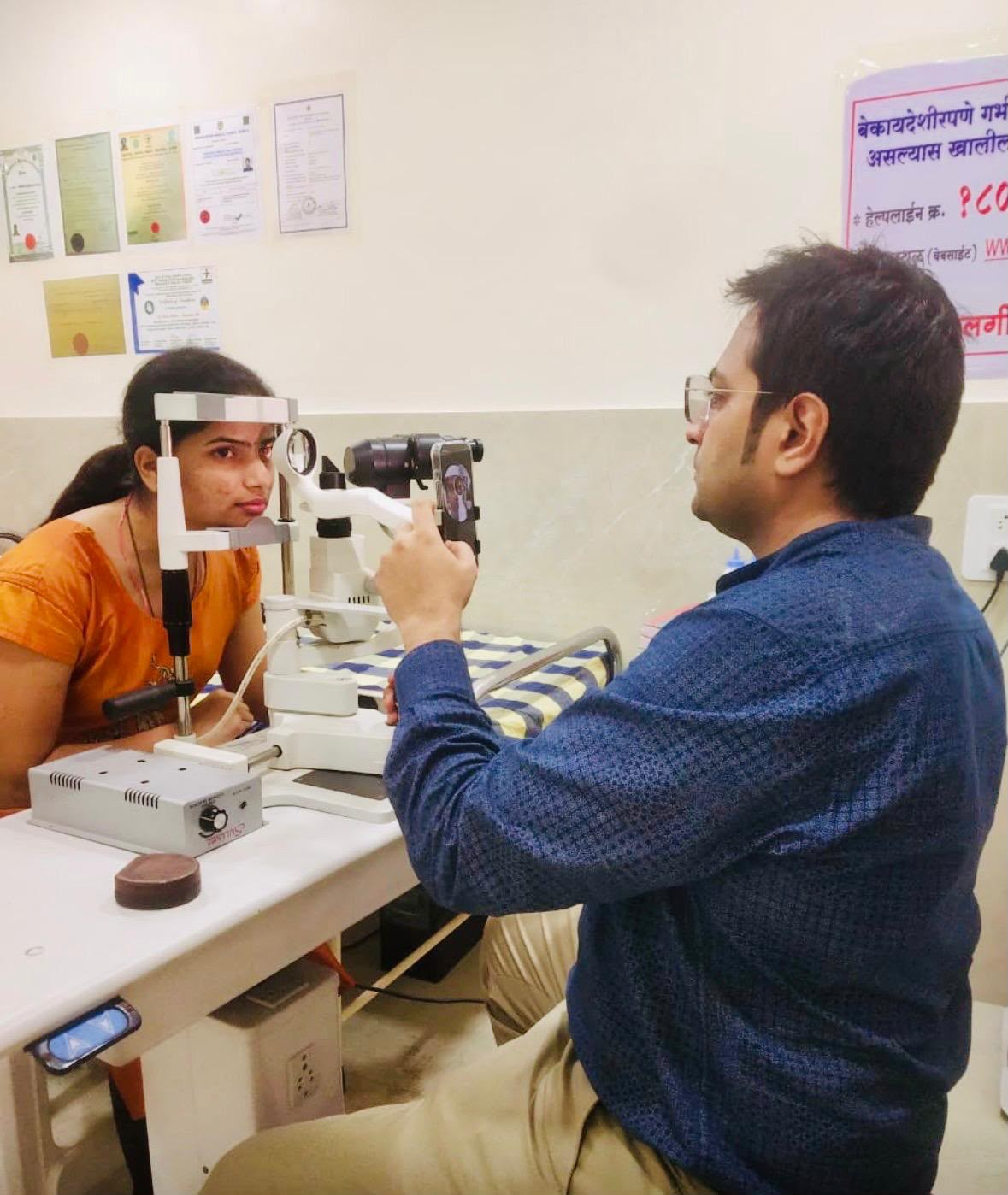
Dr. Rohit Laul
Nashik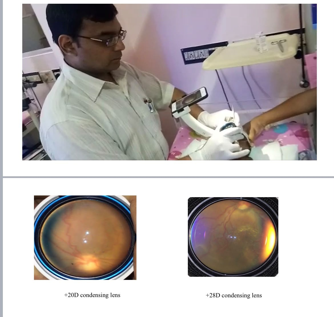
Dr. Anubhav Goyal
Giridhar Eye Institute, KochiA categorized library of anterior segment, gonio, disc, central fundus, peripheral fundus and ROP / paediatric images — all captured using MII devices in real clinical settings. Tap a category to browse, and tap any image to view it full size.
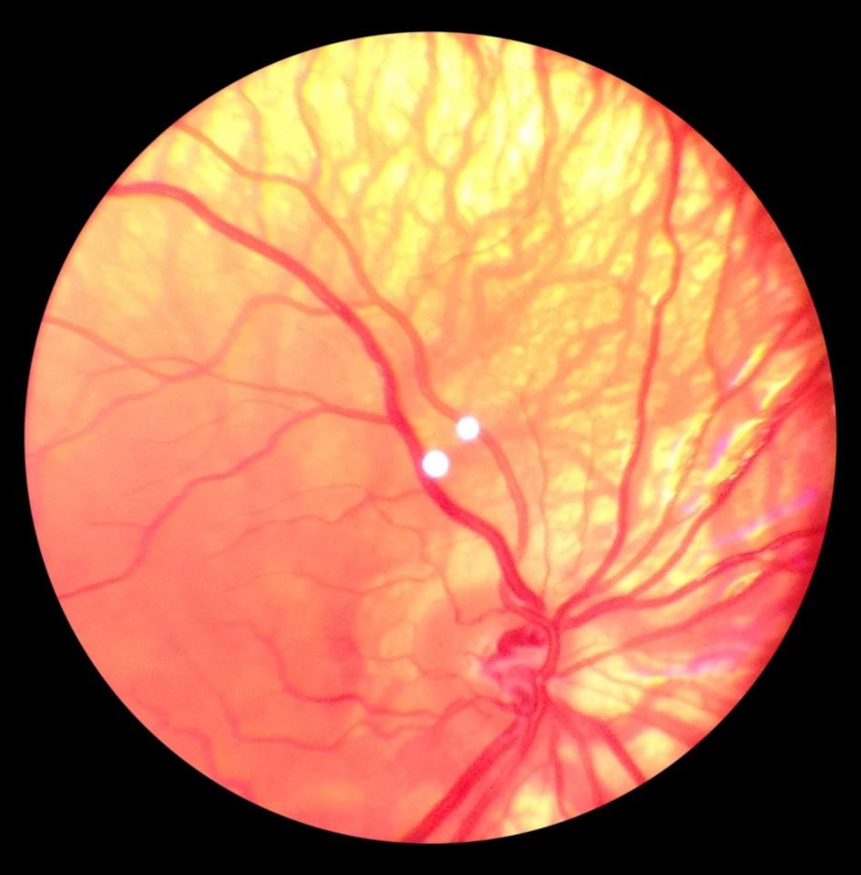 Albino Fundus
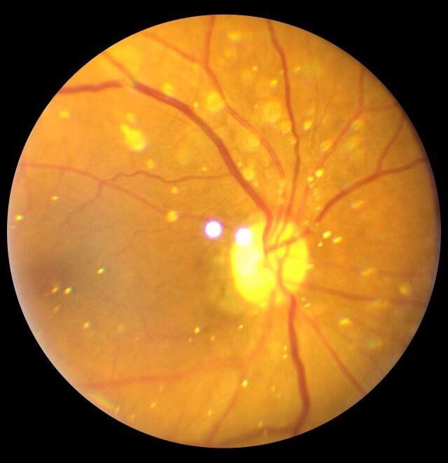 Asteroids
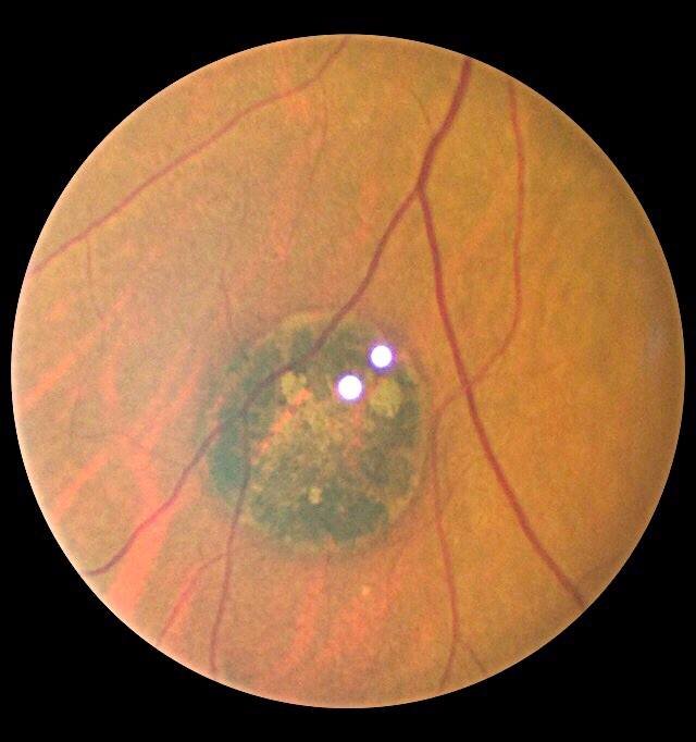 CHRPE 1
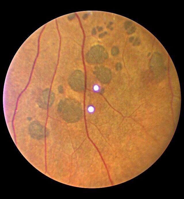 CHRPE
CHRPE
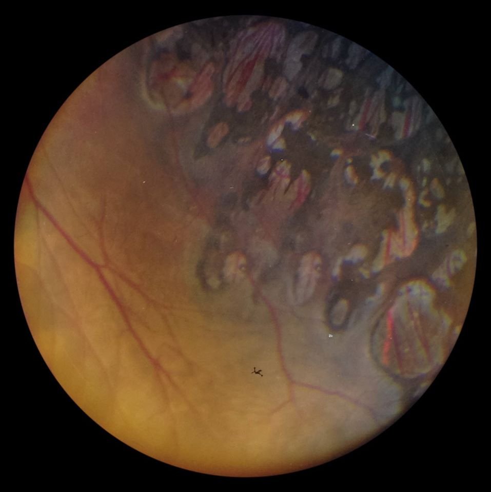 CRA
CRA
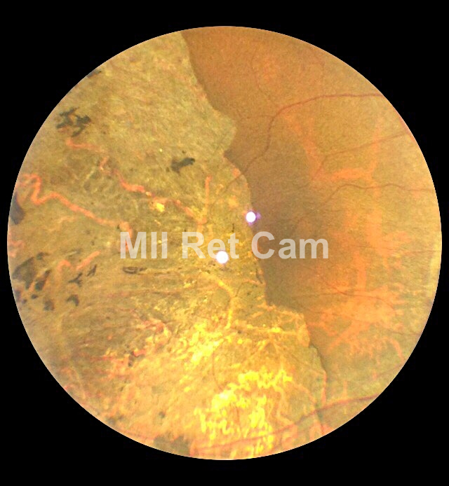 CRA
CRA
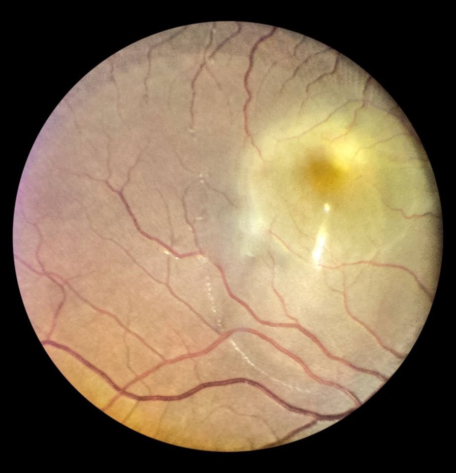 CSR 1
CSR 1
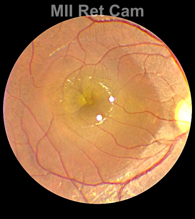 CSR
CSR
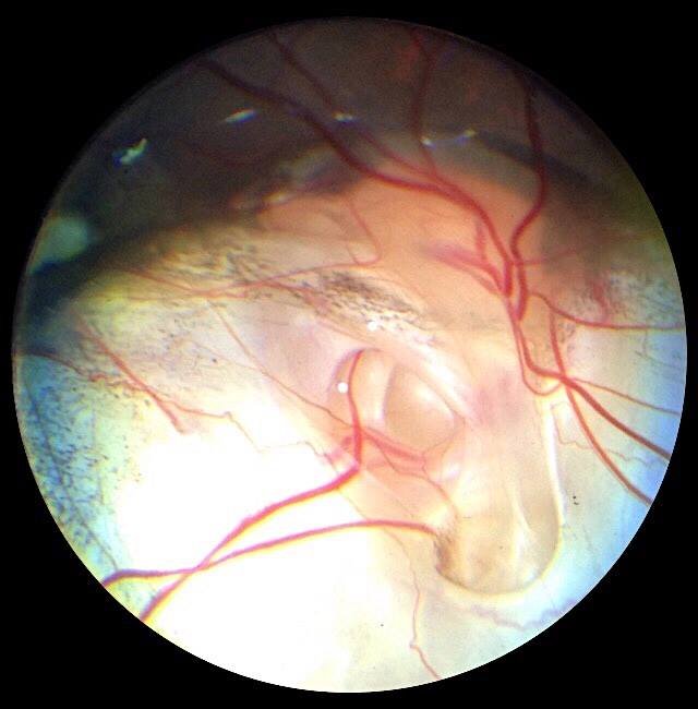 Choroidal Coloboma
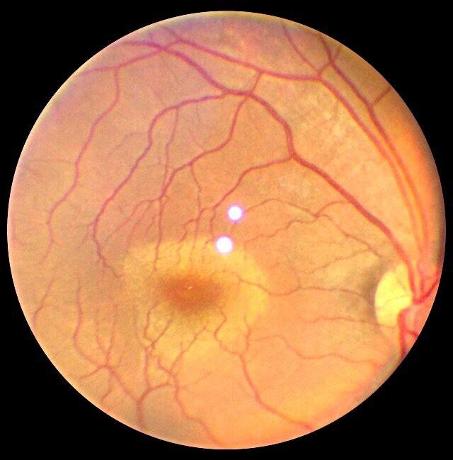 Commotio
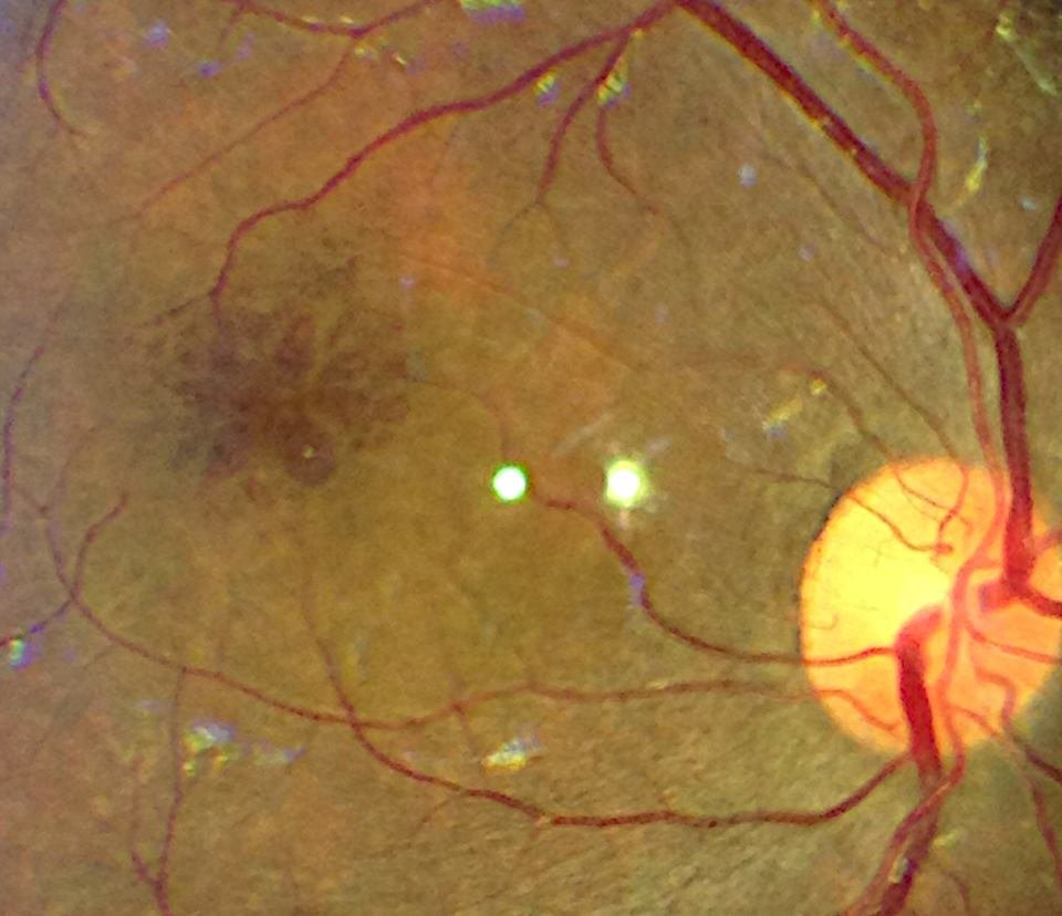 Cystoid Macular Edema
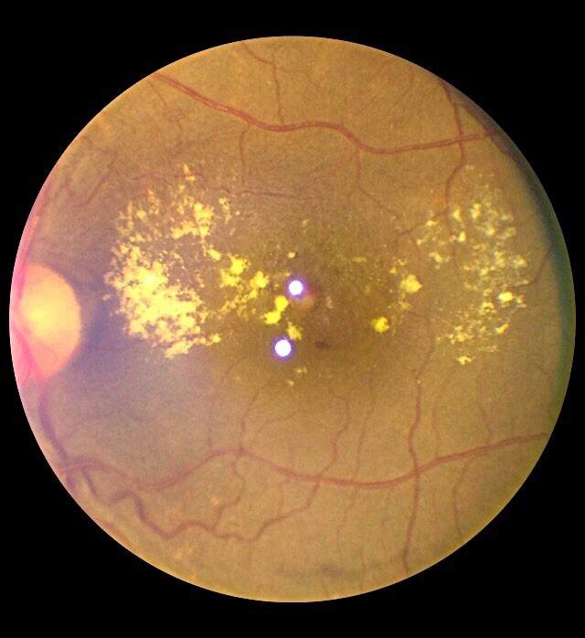 DR
DR
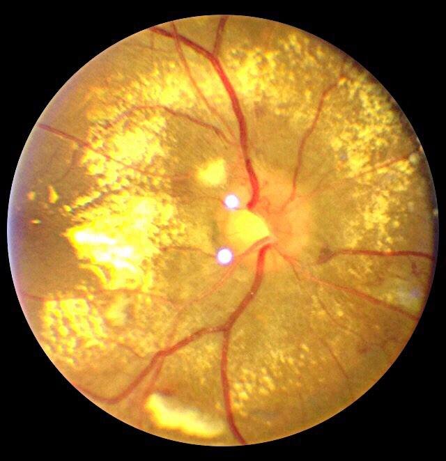 Diabetic Retinopathy
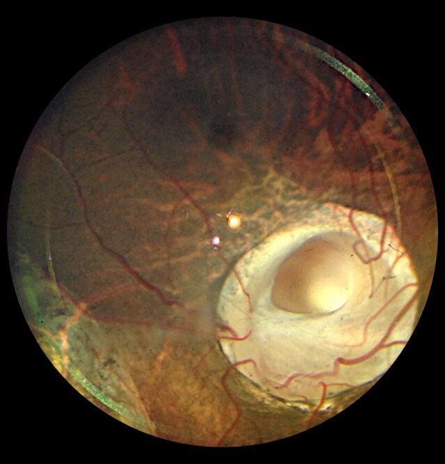 Disc Coloboma
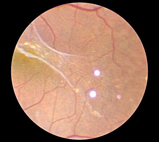 ERM
ERM
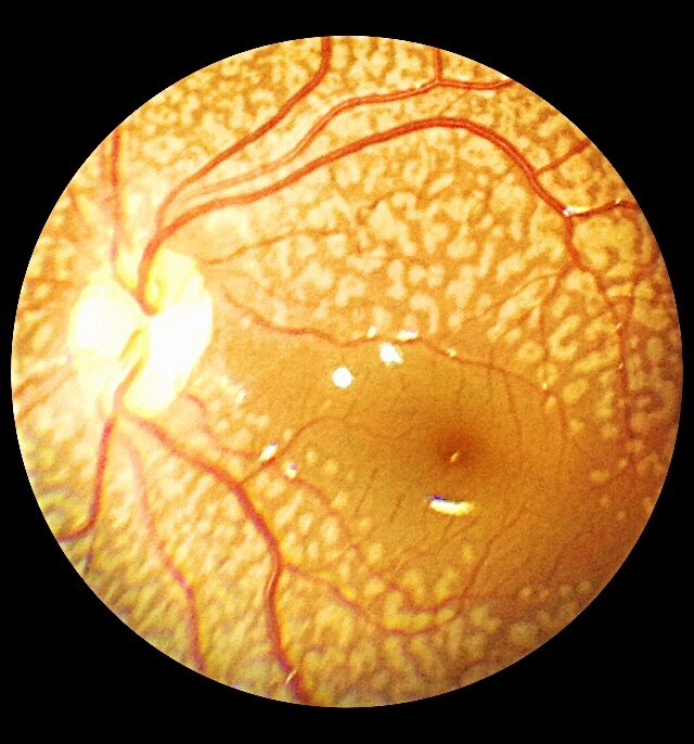 Fundus Flavimaculatus
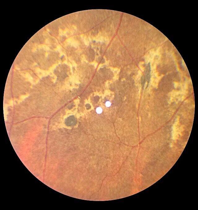 Healed Choroiditis 1
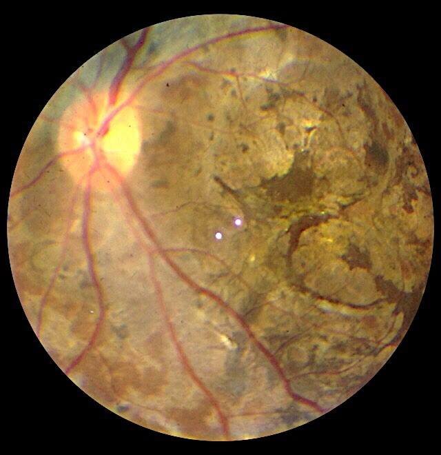 Healed Choroiditis
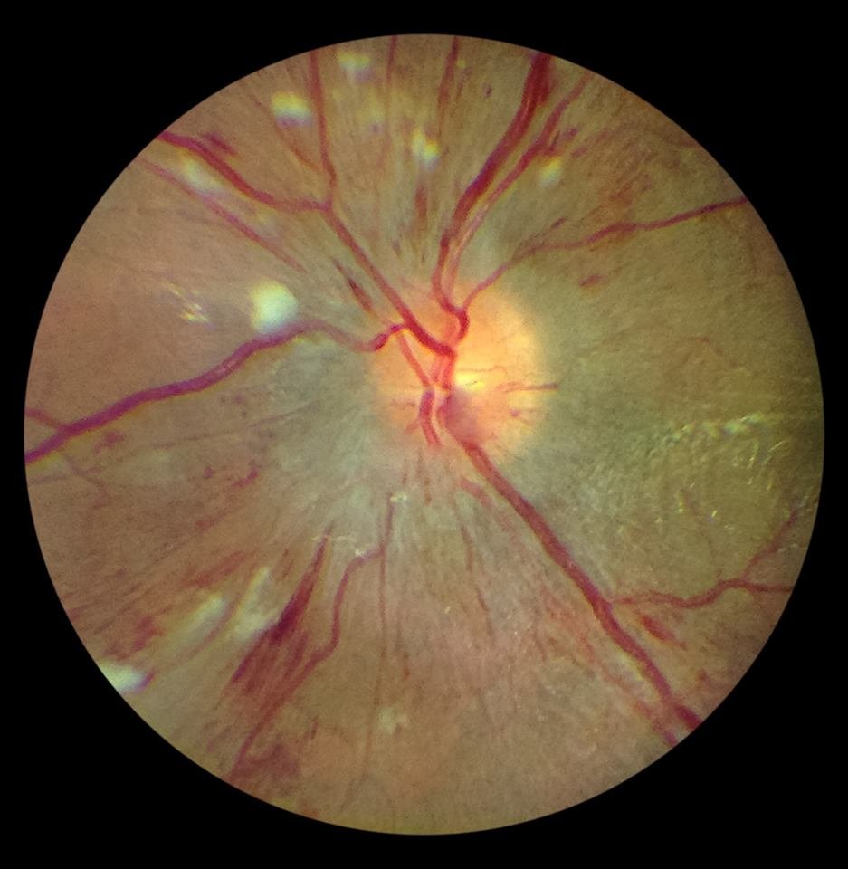 Hypertensive Retinopathy
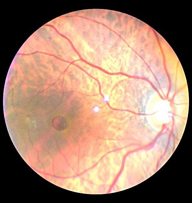 Macular Hole 1
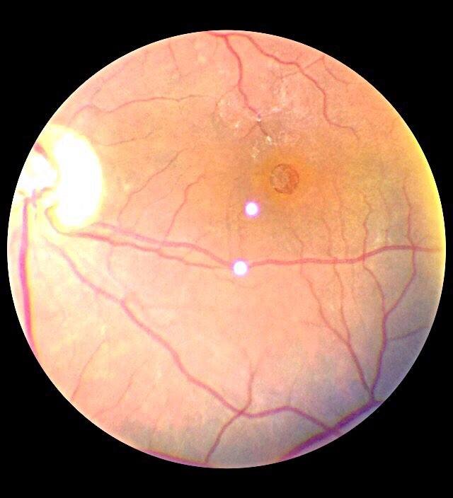 Macular Hole
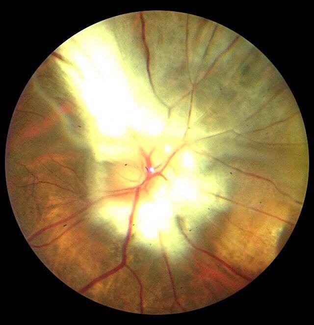 Mylinated Nerve Fibres
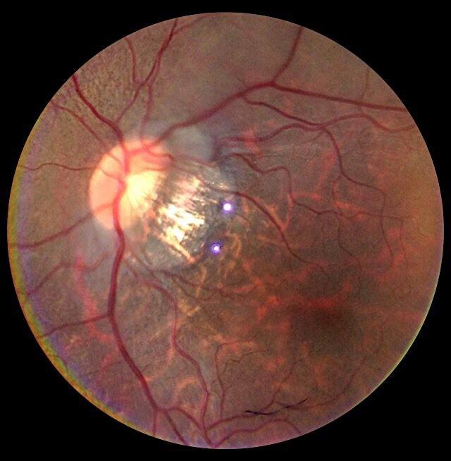 Myopic Fundus
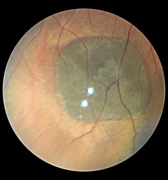 Nevus
Nevus
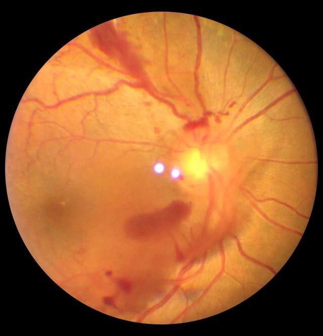 PDR
PDR
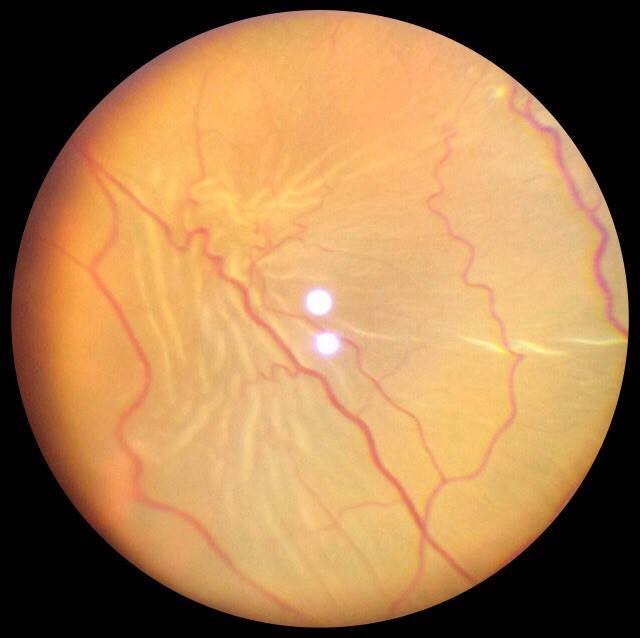 PVR
PVR
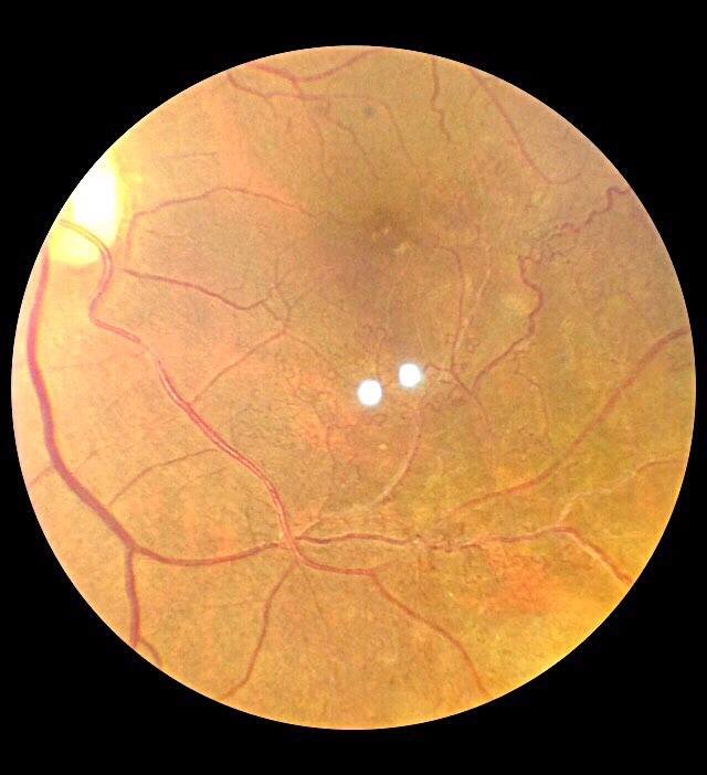 Perfused Vein Occlusion
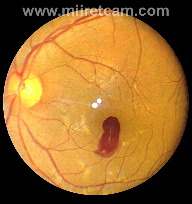 Pre retinal Hemorrhage
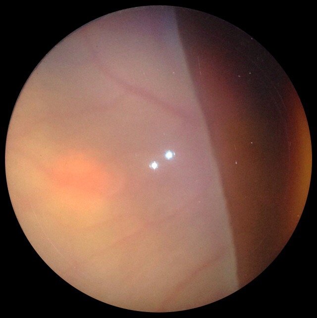 Retinal Detachment
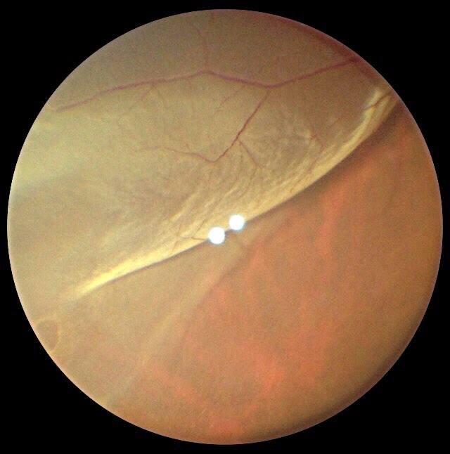 Retinal Detachment
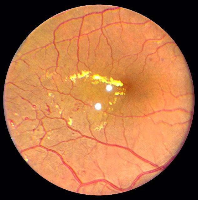 Telangiectic Vessels 1
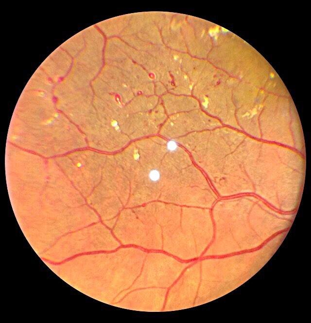 Telangiectic Vessels
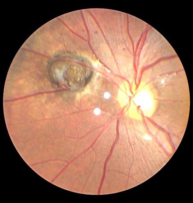 Toxo
Toxo
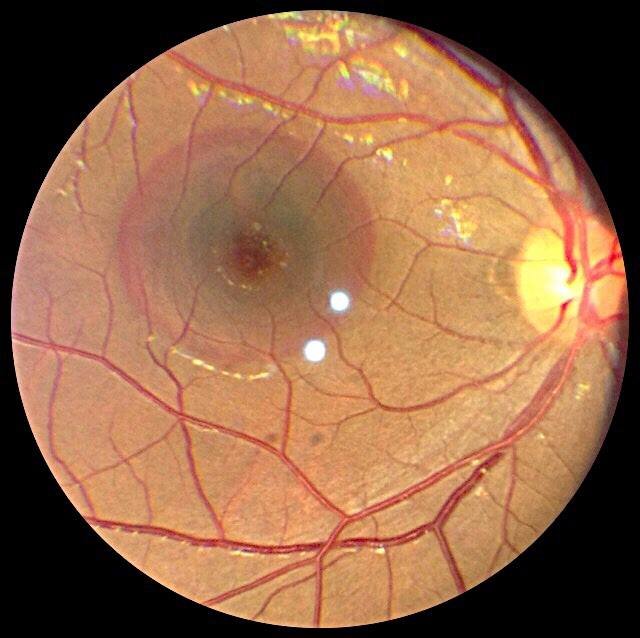 Traumatic PED
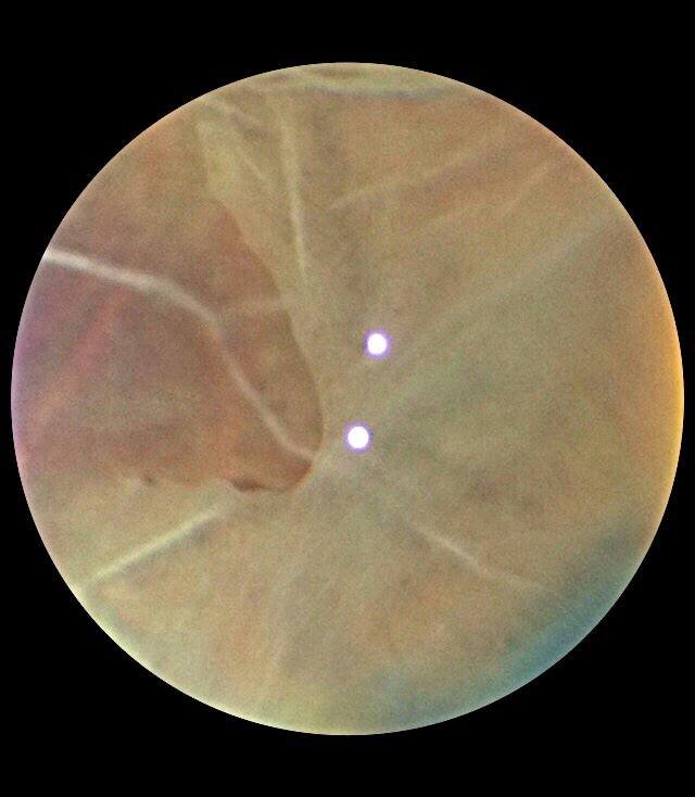 Vitreous Degeneration 1
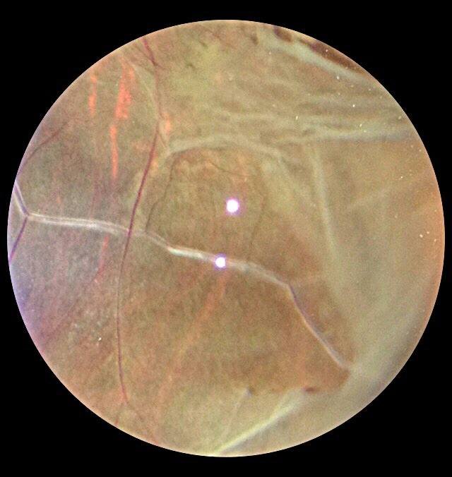 Vitreous Degeneration
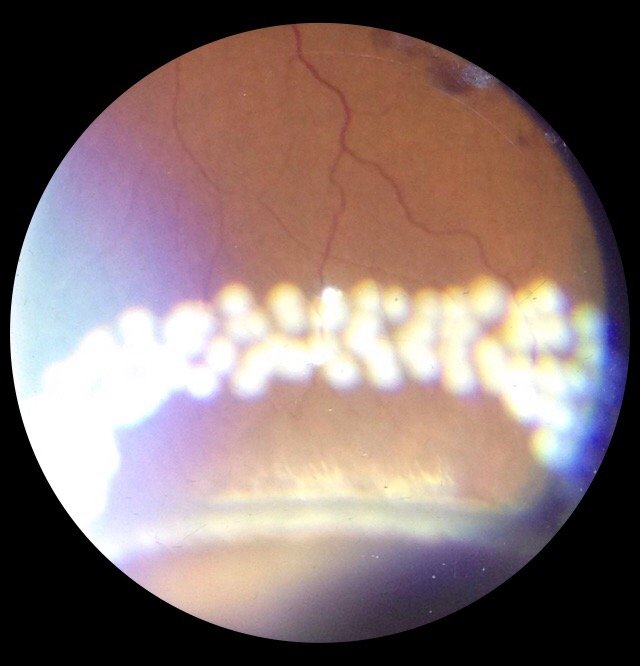 Barrage Laser
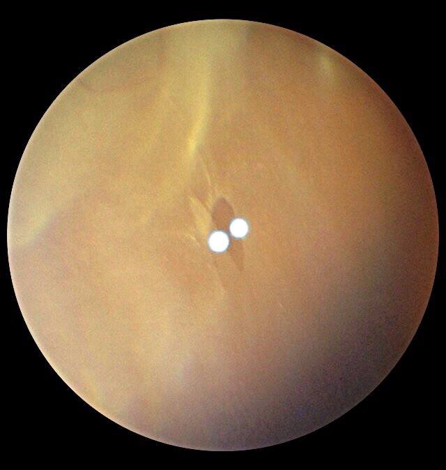 HST
HST
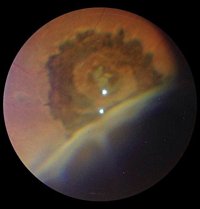 Lasered Hole
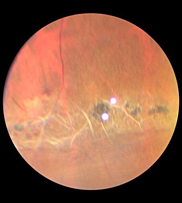 Lattice 1
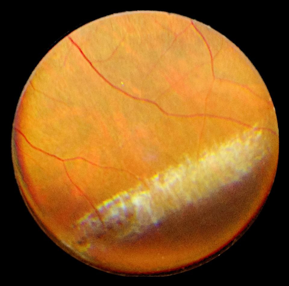 Lattice 2
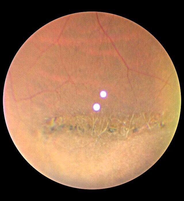 Lattice
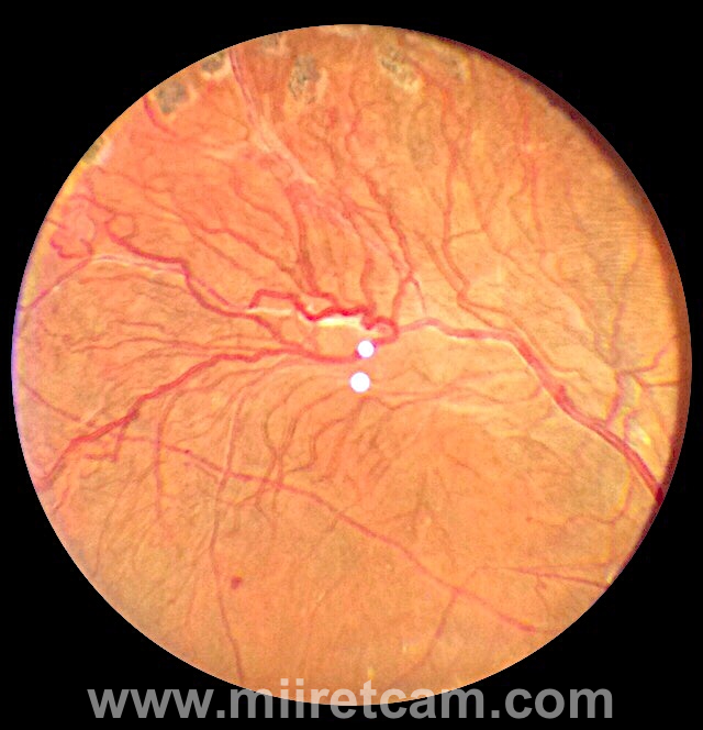 Mid Periphery
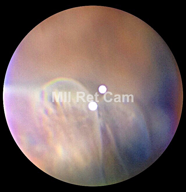 Pars Plana Cyst
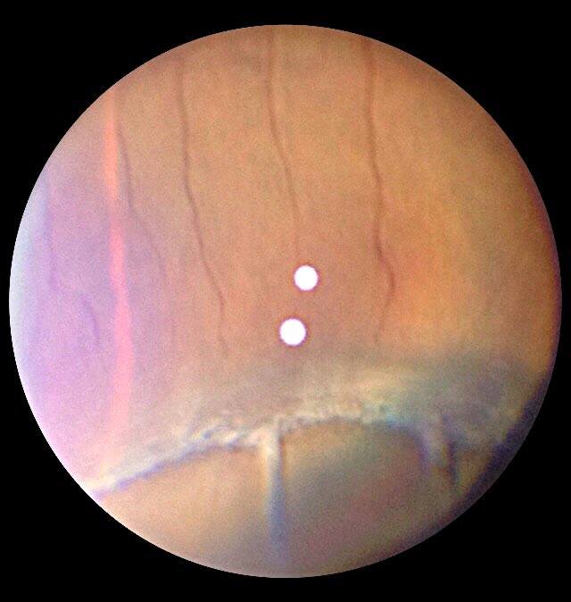 Pars Plana, Ora Serrata and Dentate Processes
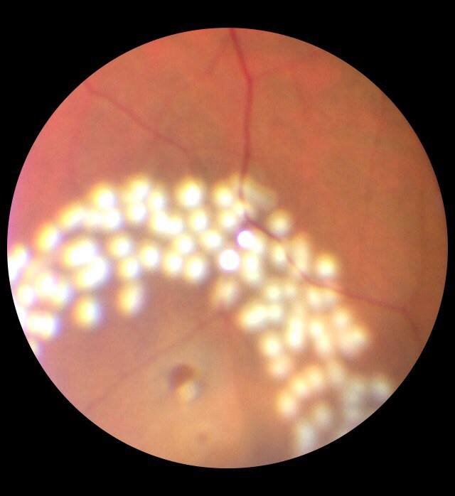 Post Barrage Hole
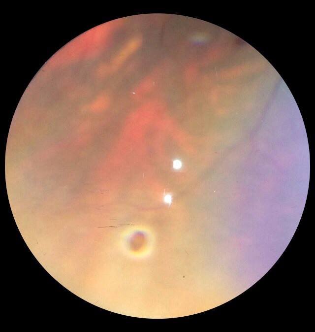 Retinal Hole 1
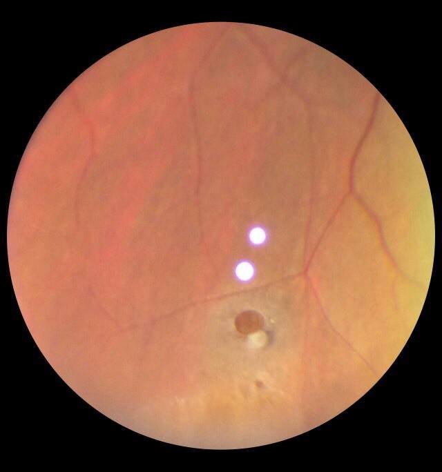 Retinal Hole with Operculum
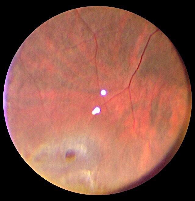 Retinal Thinning with hole
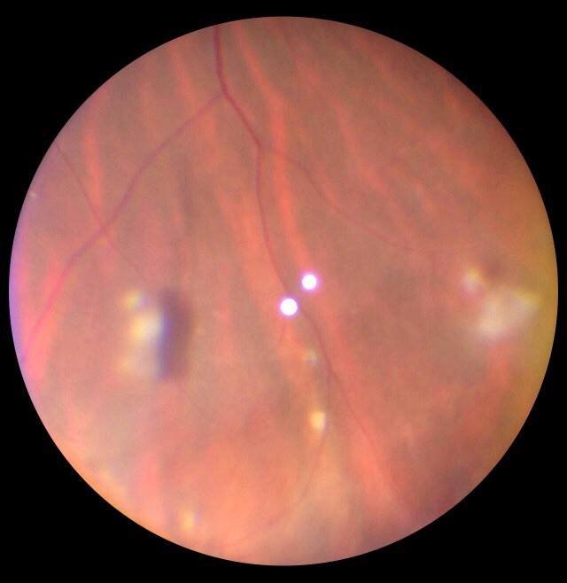 Snow Balls Intermediate Uveitis
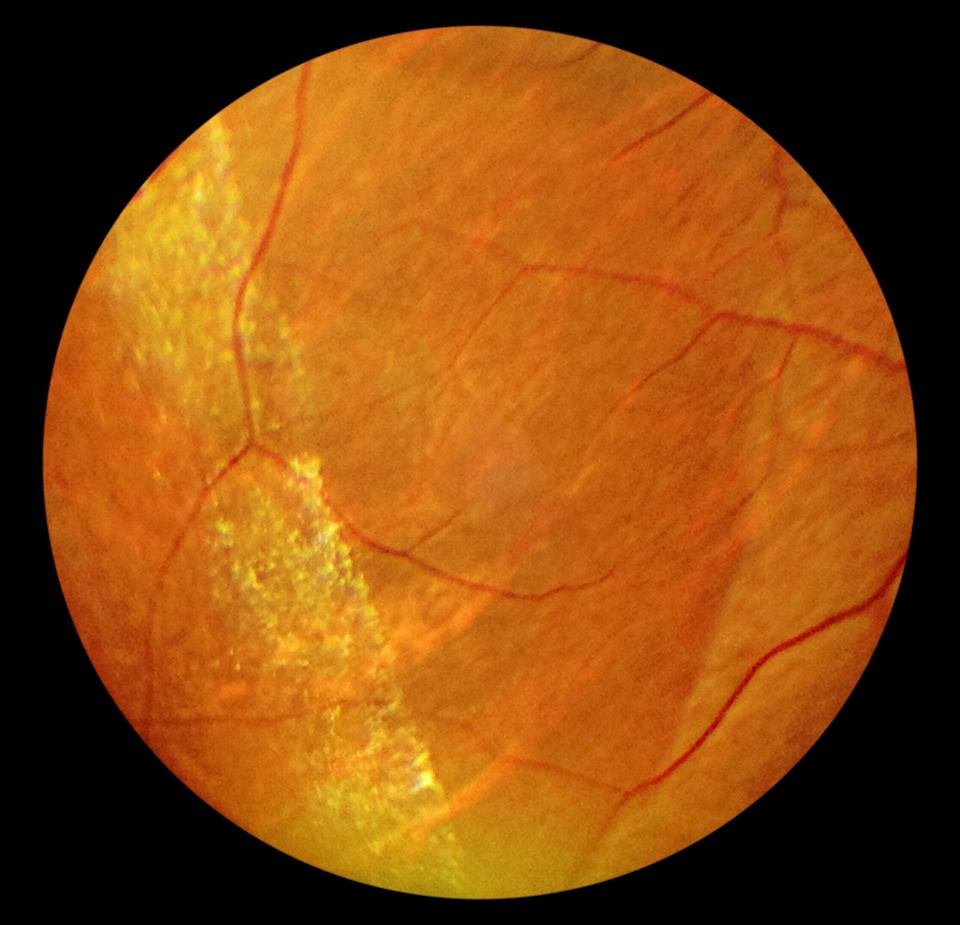 Snow Flake Degeneration
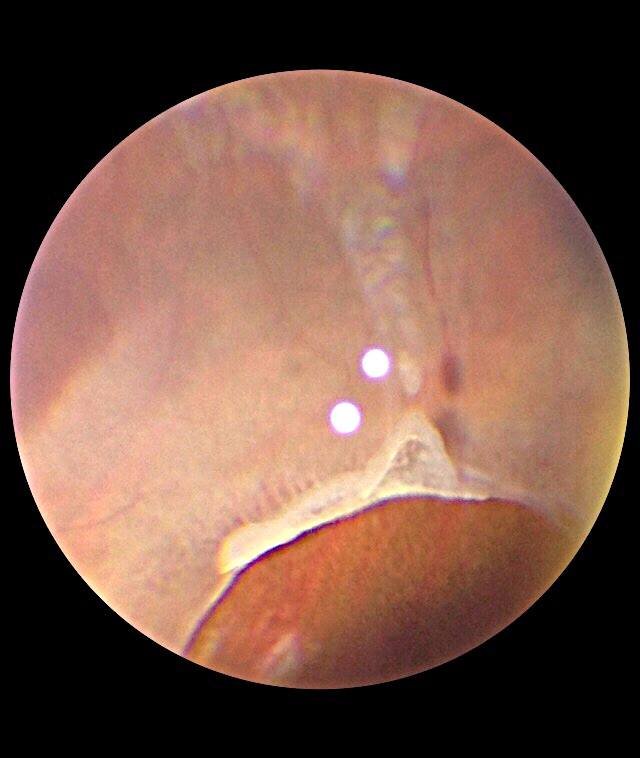 Vitreous Traction with Tear
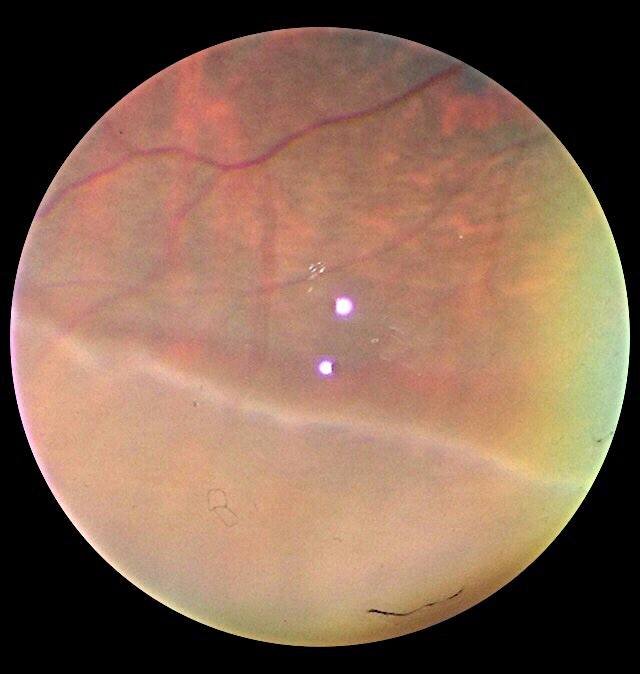 WWOP
WWOP
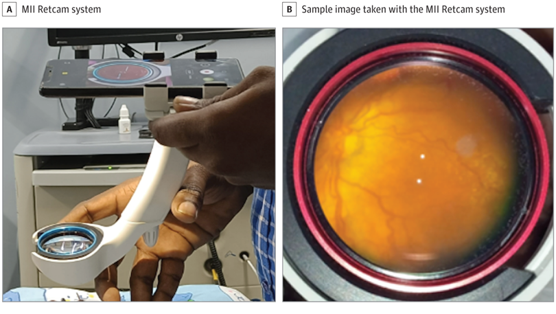 Published Image (JAMA) Courtesy -Dr Peter Cambell and Dr Shah
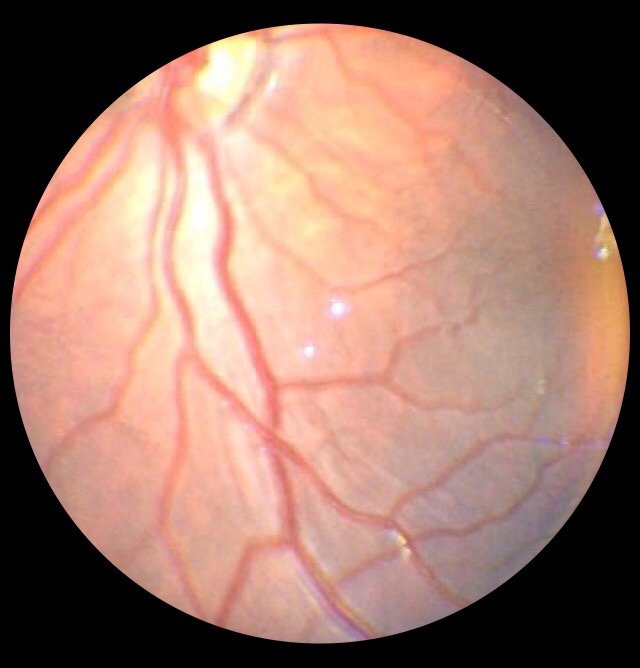 ROP 1 (8)
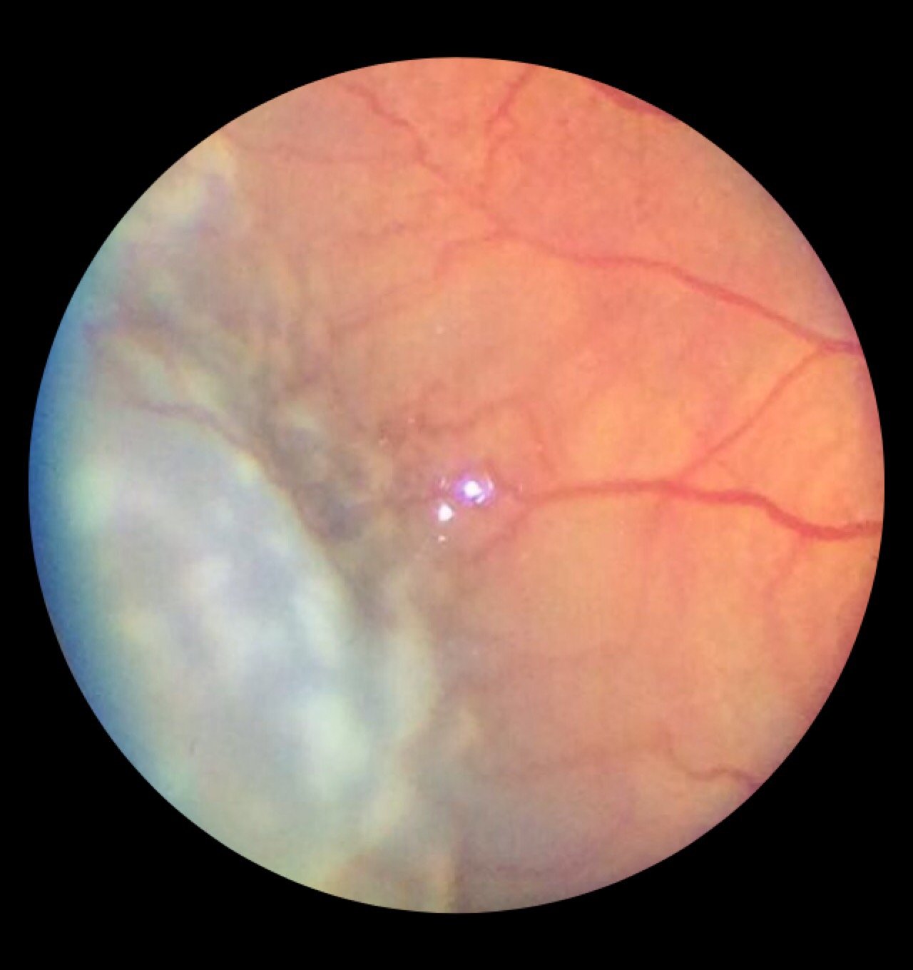 ROP 1
ROP 1
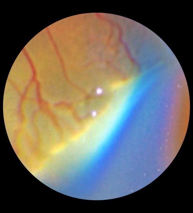 ROP 2
ROP 2
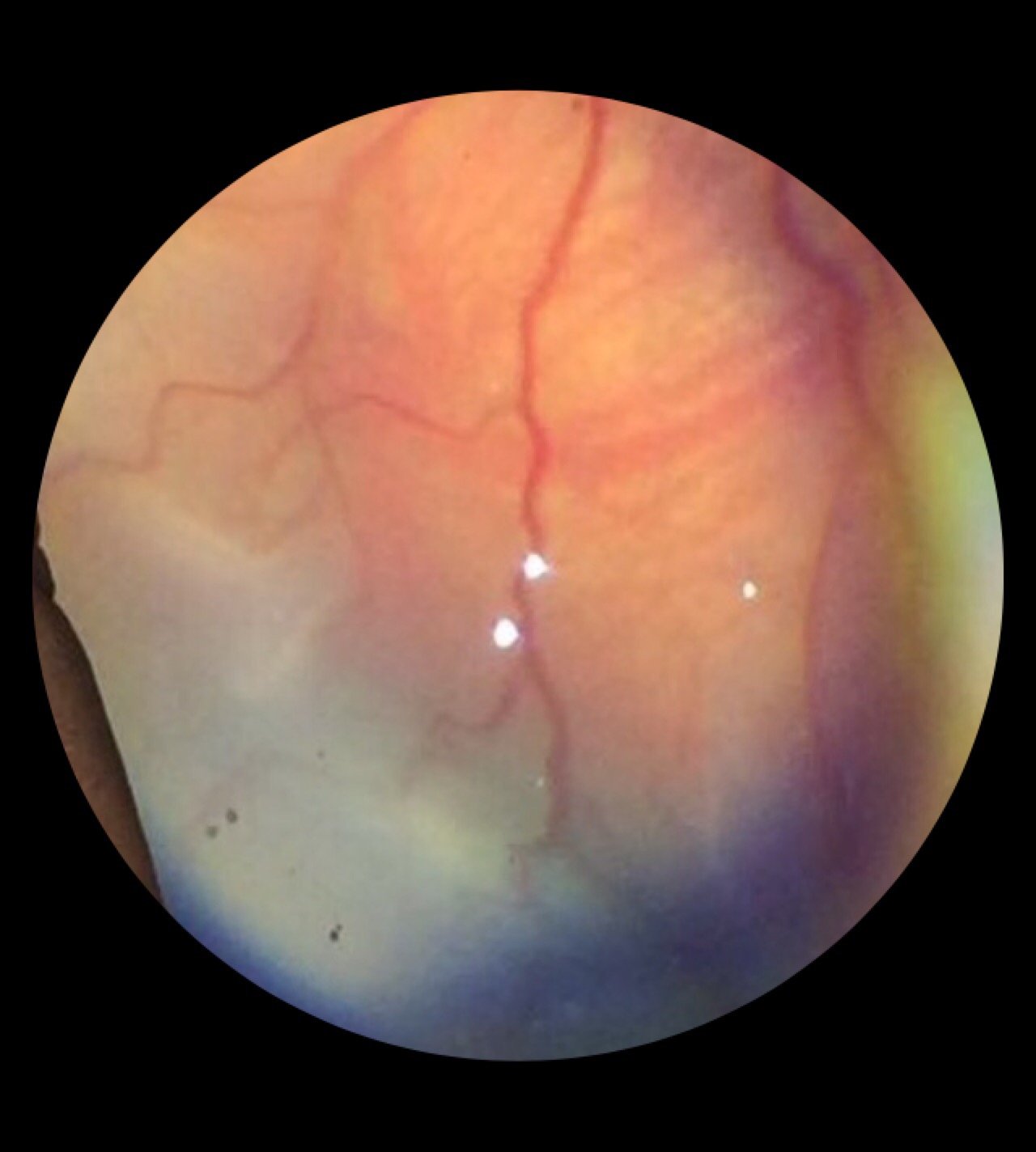 ROP 3
ROP 3
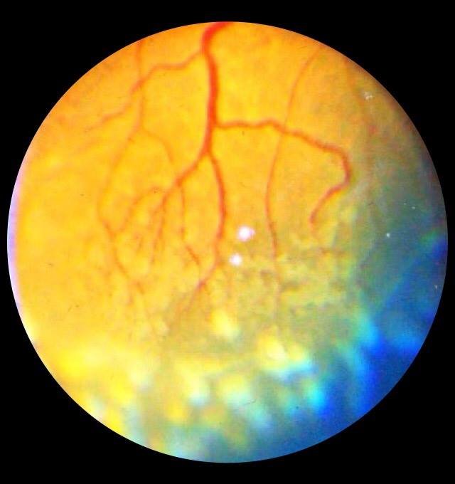 ROP 4
ROP 4
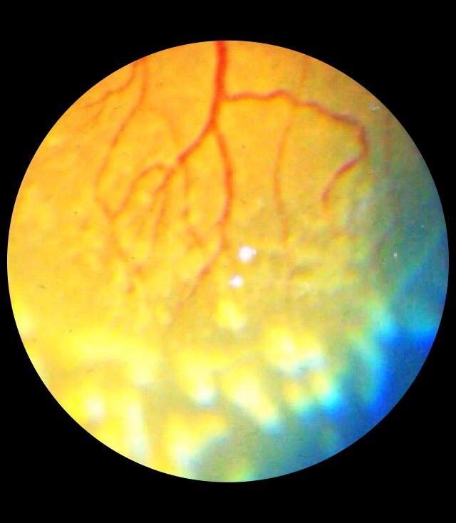 ROP 5
ROP 5
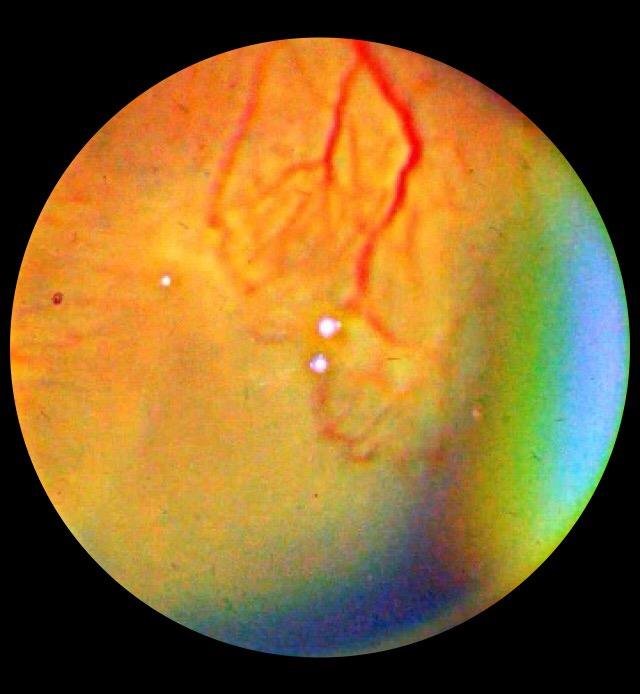 ROP 6
ROP 6
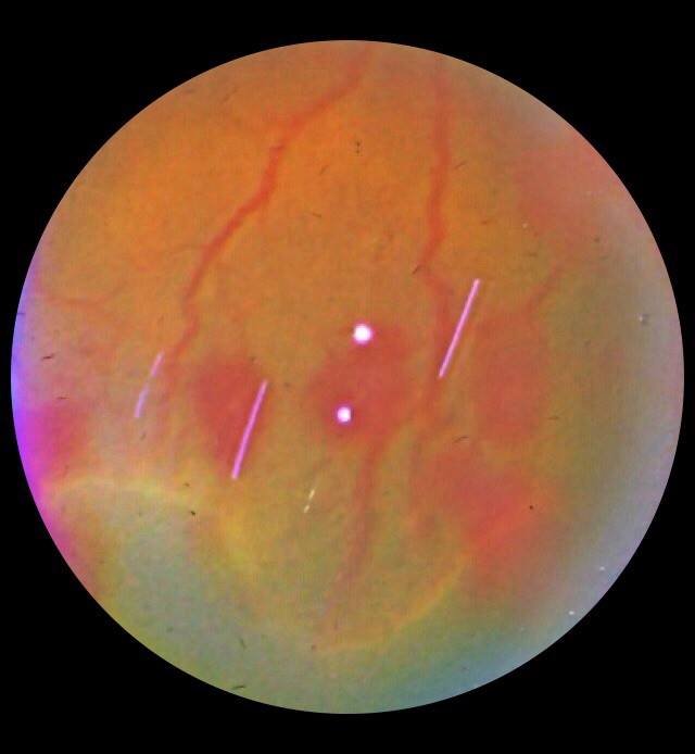 ROP 7
ROP 7
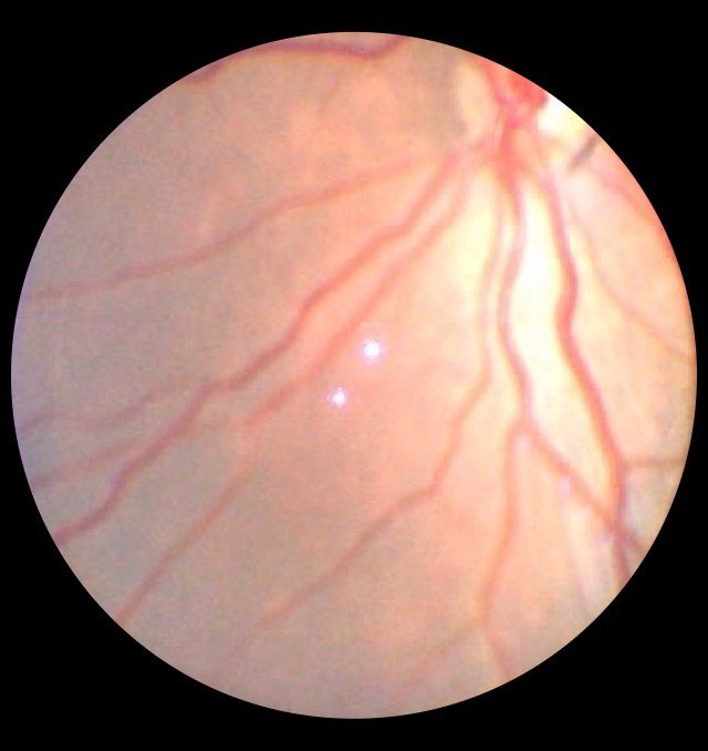 ROP 8
ROP 8
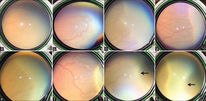 ROP Published Image (IJO) Courtesy -Dr Lekha
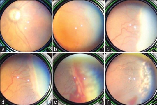 ROP Published Image 2(IJO) Courtesy -Dr Lekha
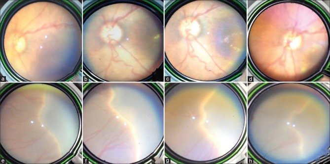 ROP Published Image 3 (IJO) Courtesy -Dr Lekha
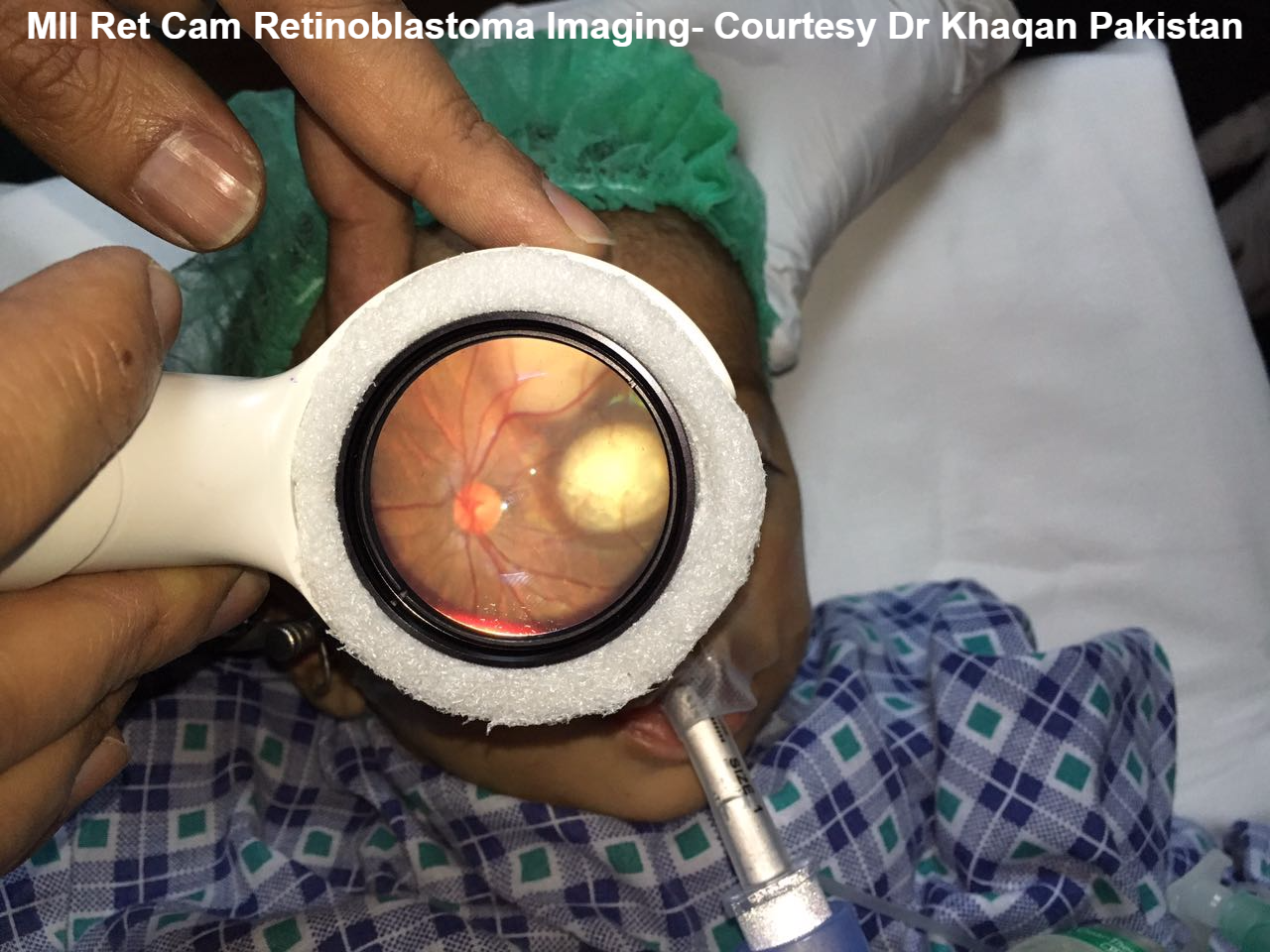 Retinoblastoma (Courtesy Dr Khaqan Pakistan)
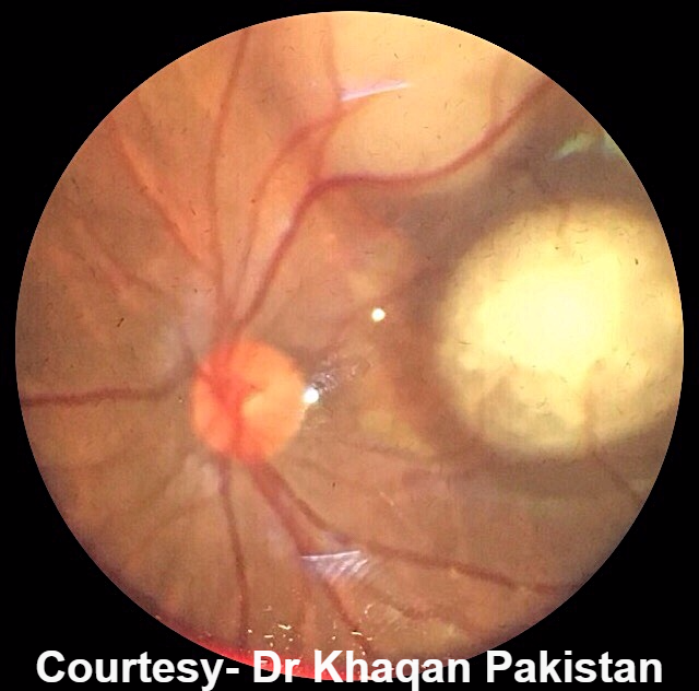 Retinoblastoma 6 (Courtesy Dr Khaqan Pakistan)
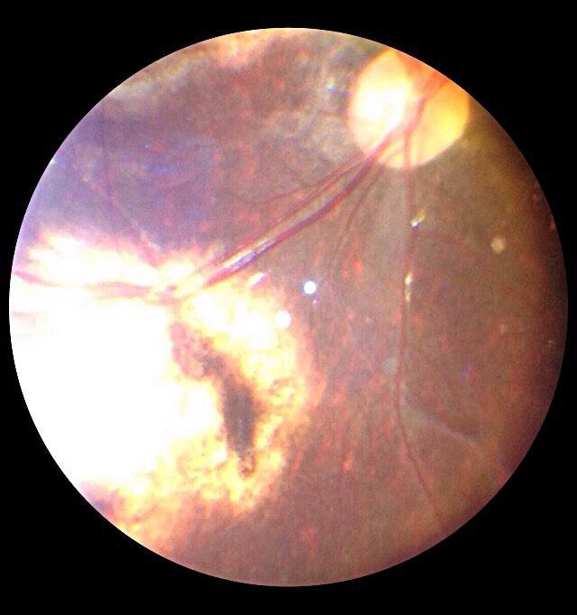 Retinoblastoma 1 (Courtesy Dr Khaqan Pakistan)
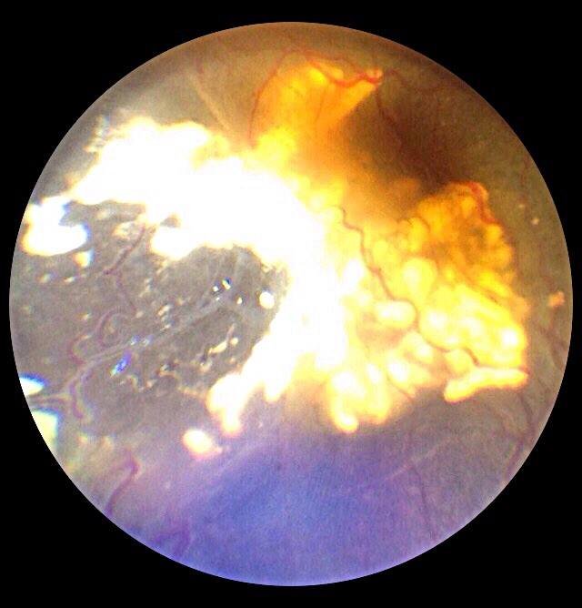 Retinoblastoma 2 (Courtesy Dr Khaqan Pakistan)
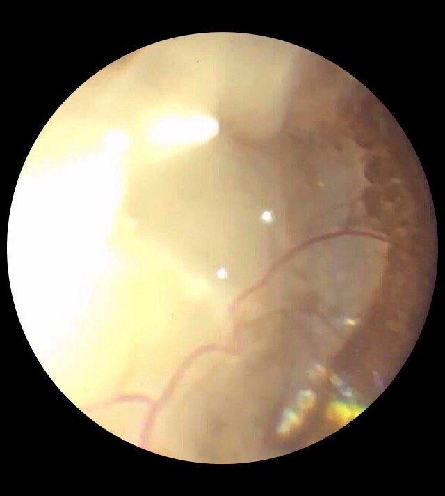 Retinoblastoma 3 (Courtesy Dr Khaqan Pakistan)
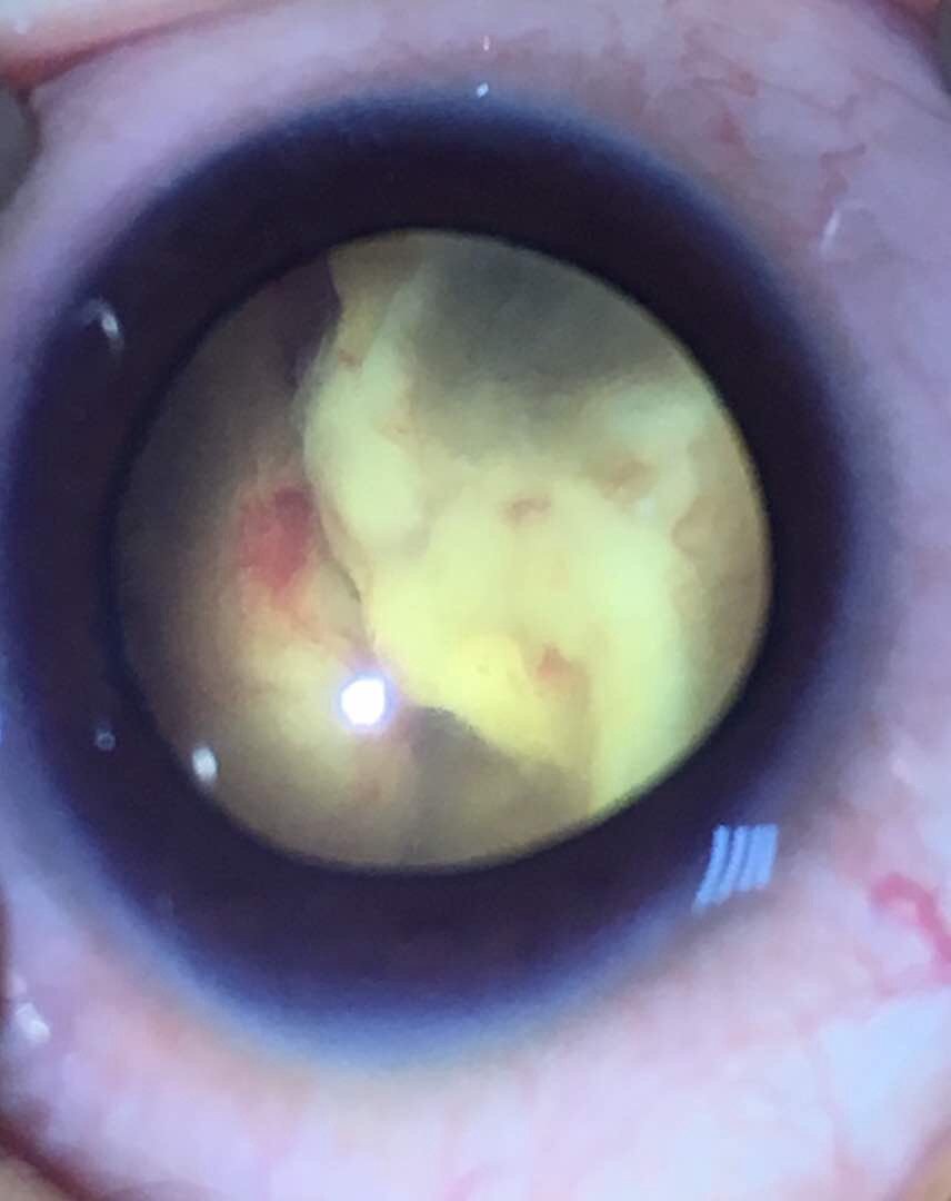 Retinoblastoma 4 (Courtesy Dr Khaqan Pakistan)
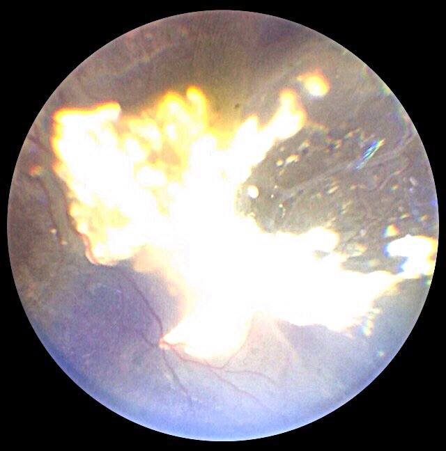 Retinoblastoma 5 (Courtesy Dr Khaqan Pakistan)
If you’ve published research using an MII device, let us know and we’ll add it here.
Share Your Research