An Idea. A Smartphone. And a Lot of Experimentation.
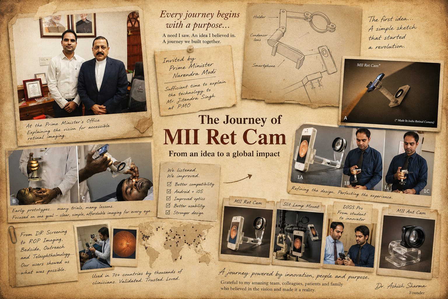
We decided to harness something that was already in everyone's hands — the smartphone. What followed was a journey of sketches, optical experiments, prototypes, failures, modifications and repeated testing.
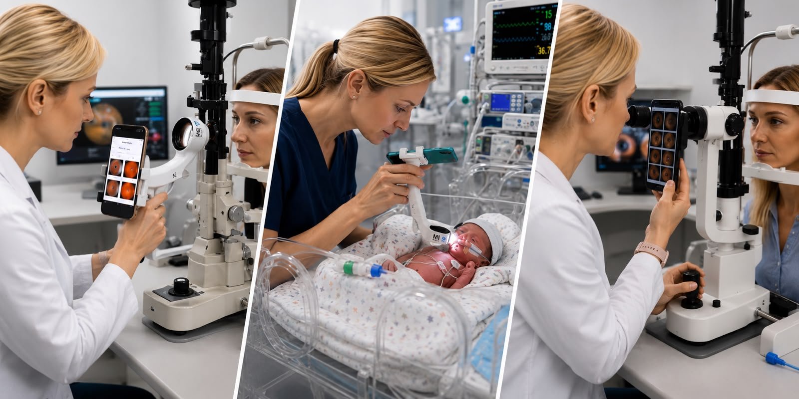
The first MII Ret Cam was designed as a simple handheld smartphone-based retinal imaging device. The philosophy was straightforward: make retinal imaging accessible wherever the patient is.
And Then the Users Took It Somewhere We Had Not Imagined
Initially, our focus was retinal imaging and diabetic retinopathy screening. But once clinicians started using MII Ret Cam, something unexpected happened. Users began using it for ROP imaging, bedside imaging and situations where bringing a conventional fundus camera to the patient was simply not practical.
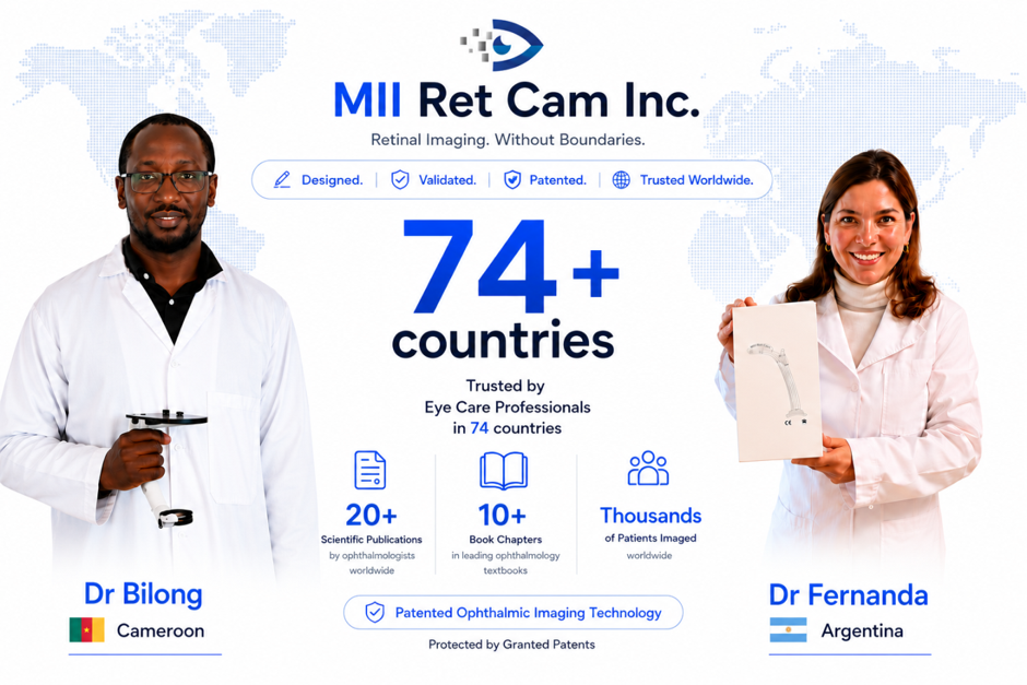
ROP imaging, in particular, opened an entirely new chapter. The device began to be used by clinicians around the world, and its clinical utility was supported by contributions and validation from global users and scientific publications. The users were not just adopting MII Ret Cam — they were helping us discover what it could become.
Listening. Learning. Improving.
Our first year was especially meaningful. With a humble beginning and without an established distributor network, MII Ret Cam began finding users across different parts of the world. Its portability, affordability and usability helped it grow organically.
But as the user base expanded, so did the feedback. During the second and third years, clinicians began telling us about limitations with certain Android devices. We listened, went back to the drawing board, and redesigned the system to improve compatibility across both Android and iOS platforms.
That became an important lesson for us: a medical device is never truly finished. It evolves with the people who use it.
From Retina to Every Eye
As MII Ret Cam reached more clinicians, we noticed something else — it was no longer being used only by retina specialists. Anterior segment specialists and optometrists were beginning to use it as well. But they wanted greater stability.
That feedback led to the MII Ret Cam Slit Lamp Mount. The objective was not to create another complicated accessory; it was to make imaging stable, intuitive and easy to perform, even without extensive training. The handheld device could now move from bedside to slit lamp with ease.
And again, the users surprised us. MII Ret Cam began finding applications beyond ophthalmology — with neurologists, pediatricians, physicians, gynecologists and even veterinarians exploring its possibilities. What began as a retinal imaging device was becoming a broader clinical imaging platform.
A Fellow's Idea Changed the Journey Again
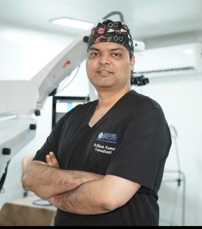
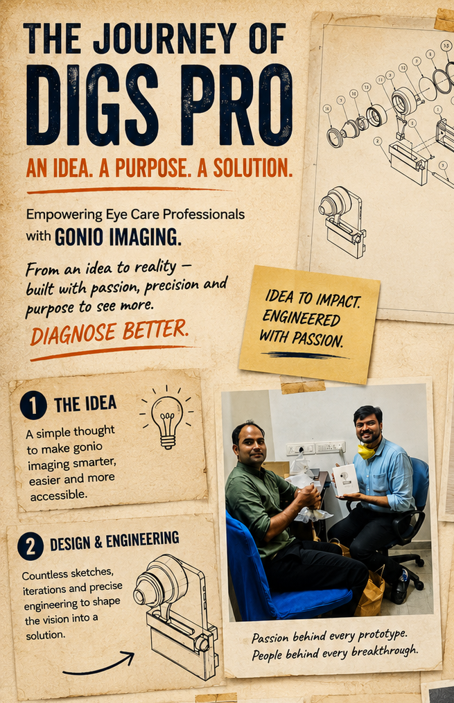
One of the most rewarding parts of this journey has been seeing young clinicians contribute ideas that take an innovation in an entirely new direction. My fellow, Dr. Nilesh Kumar, came up with an idea that challenged us: could we image the anterior chamber angle using a smartphone?
At the time, this was not something readily available as a smartphone-based solution. We worked on the concept together and developed what we believe to be the world's first smartphone-based angle imaging device — a reminder that innovation often comes from simply asking, “Why not?”
MII Ant Cam
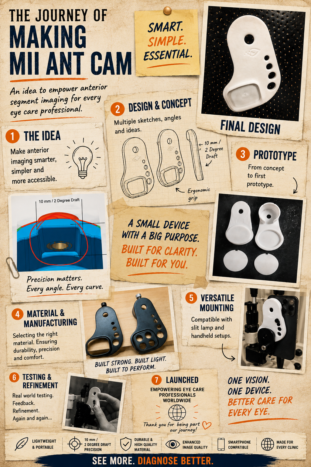
As our community of users grew, another question kept coming back to us: “Do you have something for anterior segment imaging?” We looked at what was available and found that many existing smartphone adapters were relatively complex and not specifically designed around the needs of everyday anterior segment imaging.
So we went back to research and development. The result was MII Ant Cam — a compact, magnetic, screw-free system designed to make anterior segment imaging simple and practical. The response was beyond what we expected: more than 100 ophthalmologists booked MII Ant Cam even before its official launch, and the feedback since has reinforced what we've believed from the beginning — simplicity can be powerful.
The Journey Is Far From Over
Today, MII is no longer just about one device. It is a journey of clinical problems, simple ideas, engineering, user feedback, scientific validation and relentless refinement. From a retinal imaging device born out of a need in India, to applications in ROP, bedside imaging, slit-lamp imaging, angle imaging and anterior segment imaging — the journey has been shaped at every step by the people who believed in the idea and used it in the real world. And we are still learning. Our goal remains the same: to simplify ophthalmic imaging, make it more accessible, and explore new possibilities with every generation.
The People Behind the Journey
No innovation is ever the work of one person. MII has been built by an extraordinary team of people who contributed their time, skills, ideas and persistence at different stages of the journey. We are deeply grateful to Mr. Senthil, Mr. Shajan, Mr. Ramachandran, Mr. Shreedhar, Mrs. Saranya, Mr. Lakshmilanthan, Mr. Brijesh, our packaging designer, and Mr. Govindarajalu, who has taken responsibility for worldwide shipping.
A special thanks to our software team — Mr. Raman, Mr. Kannapiran, Mr. Abhijeet, Mr. Ravi and Mr. Ramachandran — whose work has been critical in turning our hardware innovations into practical, usable solutions. We would also like to acknowledge Mr. Raj and Mr. Anand, dear friends who supported and encouraged us throughout this journey.
We are equally grateful to our patients, who allowed us to test, learn and validate these technologies in real clinical practice; to our global colleagues and users, whose feedback has continuously challenged us to improve; and to every clinician who gave MII a chance.
Behind the Innovation Is a Family
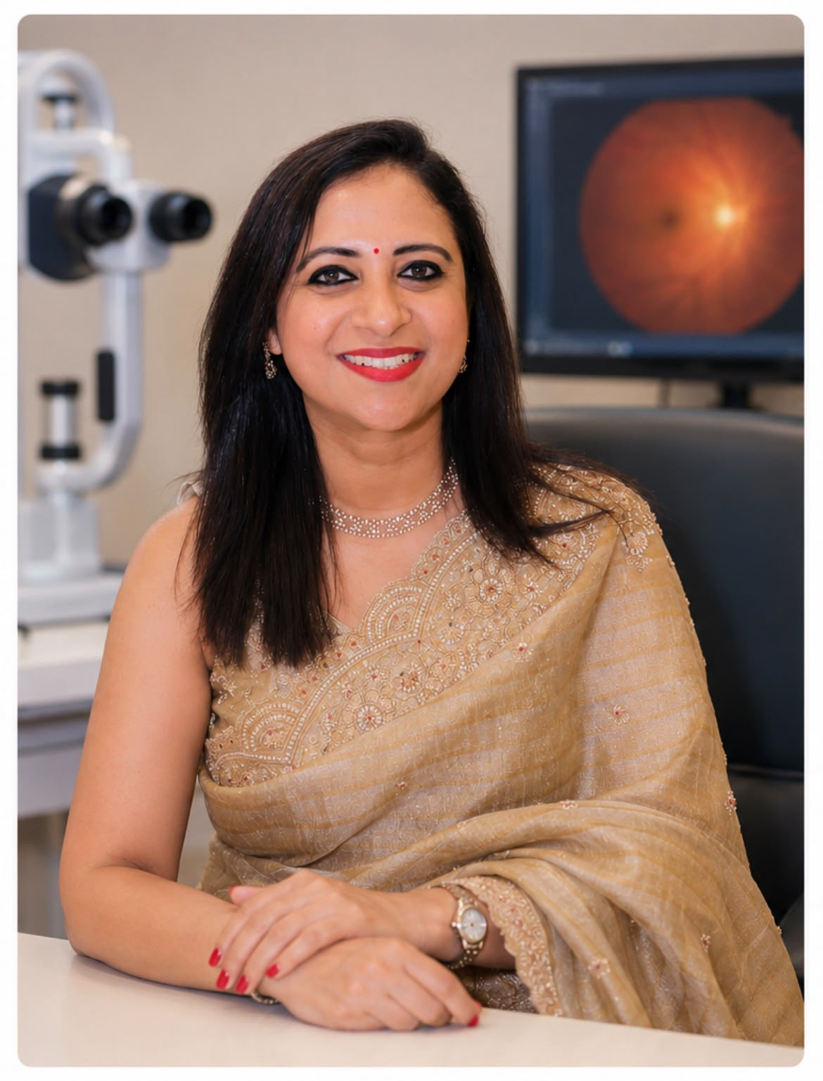
Every entrepreneurial journey has a story beyond the laboratory and the clinic. A very special thanks to Dr. Rohini Sharma, whose constant support, encouragement and guidance have been an integral part of MII's journey — her perspective, involvement and steady guidance have helped us navigate important decisions and keep the company moving in the right direction.
And to Rudraksha Sharma, thank you for being part of this journey and for understanding the time, effort and passion that MII has demanded over the years. And to our parents, whose values of hard work, persistence and perseverance shaped who we are today — thank you.